LESSON 0: PROCEDURE
ARTERIAL PUNCTURE
The first thing we should know in
the field of research is that a prior theoretical and practical knowledge of
the profession is necessary.
Below is the procedure for arterial
puncture discussed in the first large group class, the basis of an article and
a video of YOU TUBE about how they did this technique in an American hospital.
With the help of this material we deduce which methodology was better, to use
local anesthetics in spite of having to perform the interventions of the
patient, because a decrease of the pain was appreciated.

- Choose the puncture site: radial artery (1st), humeral (2nd) and femoral (3rd).
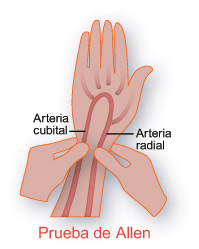
- Allen test (collateral circulation).
- Cleaning the skin with alcohol (antiseptic).
- Ask the patient if they are hypersensitive to anesthesia and if they are receiving anticoagulant treatment.
- Wear disposable gloves on the puncture with a subsequent handwash.
- Inject subcutaneously a small amount (0.3ml) of local anesthetic, which has no adrenaline.
- Check that the infiltrated area is fully anesthetized.
- Place the patient's wrist hyperextended at an approximate angle of 45 ° with the needle.
- Use needles of caliber less than 20 g.
- Ideally, a reflux of pulsatile blood should be obtained, capable of raising the plunger of the syringe passively, obtaining between 2 and 5 ml.
- Vigorously compress the puncture site for 2-3 minutes in order to prevent hematoma formation.
- Longer compression (15-20 min) may be necessary in patients with hemorrhagic diathesis.
- Ensure that the tide is watertight using the plug at the tip of the needle or similar médium.

PROCEDIMIENTO
PUNCIÓN ARTERIAL.
Lo primero que debemos saber dentro
del campo de la investigación es que es necesario un conocimiento teórico y
práctico previo de la profesión enfermera antes de disponernos a pensar sobre
qué investigar.
A continuación se expone el procedimiento de
punción arterial que comentamos en la primera clase de grupo grande, en base a
un artículo y un video de YOU TUBE sobre cómo hacían ésta técnica en un
hospital americano. Con ayuda de este material deducimos qué metodología era
mejor, utilizar anestésicos locales a pesar de tener que realizar dos
intervenciones al paciente, porque se apreciaba una disminución del dolor.
- Escoger la zona de punción: arteria radial (1ª), humeral (2ª) y femoral (3ª).
- Prueba de Allen (circulación colateral).
- Limpieza de la piel con el alcohol (antiséptico).
- Preguntar al paciente si tiene hipersensibilidad a la anestesia y si está recibiendo tratamiento anticoagulante.
- Utilizar guantes desechables en la punción con un posterior lavado de manos.
- Inyectar subcutaneamente una pequeña cantidad (0.3ml) de anestésico local, que no tenga adrenalina.
- Comprobar que la zona infiltrada se halla plenamente anestesiada.
- Colocar la muñeca del paciente hiperextendida formando un ángulo aproximado de 45º con la aguja.
- Utilizar agujas de calibre inferior a 20 G.
- En condiciones ideales, debe obtenerse un reflujo de sangre pulsátil capaz de elevar el émbolo de la jeringuilla de forma pasiva, obteniéndose entre 2 y 5 ml.
- Comprimir vigorosamente la zona de punción durante 2-3 min con objeto de prevenir la formación de hematoma.
- En pacientes con diátesis hemorrágica puede ser necesaria una compresión más prolongada (15-20 min).
- Asegurar que la jeringa sea hermética utilizando tapón en la punta de la aguja u otro medio similar.
ENLACE DEL VÍDEO: https://www.youtube.com/watch?v=07PzsH90iJM.
No hay comentarios:
Publicar un comentario